lv segments|lv segments echo : 2024-10-22 Segments of the left ventricle. Based on anatomical landmarks and autopsy studies (Edwards et al), the left ventricle is divided into three . Note: You can only have one Deck Buff active at one time! For example, if I hatched all Four Holy Beasts, ( Azulongmon, Ebonwumon, Zhuqiaomon and Baihumon ), I can have a certain effect as below:: Activation condition: Always Active. Activation Effect: Critical hit damage 30% /25% increase.
0 · myocardial segments
1 · lv wall segments echo
2 · lv segments echo
3 · coronary artery segments
4 · 17 wall segments echo
5 · 17 segments of the heart
6 · 17 segments of left ventricle
7 · 16 segment lv model
I was told this by a person working in a ford dealership parts department when I asked for Mercon SP transmission fluid. He informed me that SP was replaced by LV for 6R60/80 transmission. I have also seen this confirmed by FordTechMakuloco and others. Below is a video where he says to use LV in 6R60/80 transmissions.
lv segments*******Segments of the left ventricle. Based on anatomical landmarks and autopsy studies (Edwards et al), the left ventricle is divided into three .
Segments 1, 2, 7, 8, 13, 14, and 17 are assigned to the left anterior descending coronary artery distribution. Segments 3, 4, 9, 10, .lv segments echoLV segmental anatomy. The regional distribution of myocardial ischemia can be detected as segmental LV wall motion abnormalities by TEE. The entire LV in the 17-segment model .
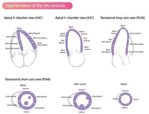
Segments of the left ventricle. Based on anatomical landmarks and autopsy studies (Edwards et al), the left ventricle is divided into three equal parts along the long axis of the ventricle. This creates three circular sections of the . Segments 1, 2, 7, 8, 13, 14, and 17 are assigned to the left anterior descending coronary artery distribution. Segments 3, 4, 9, 10, and 15 are assigned to the right coronary artery when it is dominant.LV segmental anatomy. The regional distribution of myocardial ischemia can be detected as segmental LV wall motion abnormalities by TEE. The entire LV in the 17-segment model can be imaged in long-axis using a combination of the TEE mid-esophageal four-chamber view (a), TEE mid-esophageal two-chamber view (b), and TEE mid-esophageal long-axis .Although certain variability exists in the coronary artery blood supply to myocardial segments, segments are usually attributed to the three major coronary arteries. Visual Assessment Semi quantitative wall motion score (1-4) can be assigned to each segment to calculate the LV wall motionAssessment of left ventricular systolic function has a central role in the evaluation of cardiac disease. Accurate assessment is essential to guide management and prognosis. Numerous echocardiographic techniques are used in the assessment, each with its own advantages and disadvantages. Recently, the consensus of the American Heart Association (AHA) 21 divided the LV into 4 walls: septal, anterior, lateral, and inferior; in turn, the 4 walls were divided into 17 segments: 6 basal, 6 mid, 4 apical, and 1 segment being the apex (Figure 2).Identification and classification of left ventricular (LV) regional wall motion (RWM) abnormalities on echocardiograms has fundamental clinical importance for various cardiovascular disease.The LV is divided into 3 sections: base, mid-cavity, and apex; and further subdivided into 17-segments: 6 basal segments, 6 mid-cavity segments, 4 apical segments, and the true apex as segment 17. The 17 segments correspond to specific coronary artery territories (1) . LV wall has been divided into 17 segments for the ease of the assessment of regional function. Apical segment, also called apical cap, is the only segment without a direct relation to the LV cavity. These segments correspond to the three main branches of coronary blood supply as well.
For each of the LV segments, a time-volume curve is displayed, demonstrating maximum and minimum volumes of that specific segment during the cardiac cycle. Hypo- or non-contractile (akinetic) segments can easily be identified based on the pattern of the curve, while diseased segments have a flattened curve.
Segments of the left ventricle. Based on anatomical landmarks and autopsy studies (Edwards et al), the left ventricle is divided into three equal parts along the long axis of the ventricle. This creates three circular sections of the .lv segments Segments 1, 2, 7, 8, 13, 14, and 17 are assigned to the left anterior descending coronary artery distribution. Segments 3, 4, 9, 10, and 15 are assigned to the right coronary artery when it is dominant.
LV segmental anatomy. The regional distribution of myocardial ischemia can be detected as segmental LV wall motion abnormalities by TEE. The entire LV in the 17-segment model can be imaged in long-axis using a combination of the TEE mid-esophageal four-chamber view (a), TEE mid-esophageal two-chamber view (b), and TEE mid-esophageal long-axis .lv segments lv segments echoAlthough certain variability exists in the coronary artery blood supply to myocardial segments, segments are usually attributed to the three major coronary arteries. Visual Assessment Semi quantitative wall motion score (1-4) can be assigned to each segment to calculate the LV wall motion
Assessment of left ventricular systolic function has a central role in the evaluation of cardiac disease. Accurate assessment is essential to guide management and prognosis. Numerous echocardiographic techniques are used in the assessment, each with its own advantages and disadvantages. Recently, the consensus of the American Heart Association (AHA) 21 divided the LV into 4 walls: septal, anterior, lateral, and inferior; in turn, the 4 walls were divided into 17 segments: 6 basal, 6 mid, 4 apical, and 1 segment being the apex (Figure 2).Identification and classification of left ventricular (LV) regional wall motion (RWM) abnormalities on echocardiograms has fundamental clinical importance for various cardiovascular disease.The LV is divided into 3 sections: base, mid-cavity, and apex; and further subdivided into 17-segments: 6 basal segments, 6 mid-cavity segments, 4 apical segments, and the true apex as segment 17. The 17 segments correspond to specific coronary artery territories (1) .
Power factor min. 0.95. Dimmable. Our dimmable LVT 60 12Volts/60Watts lightech electronic low voltage halogen lighting transformers can be dimmed by low cost TRIAC dimmers. It comes with patented auto heat regulation that also automatically acts as overload protection.
lv segments|lv segments echo